3nethra Classic HD, Non-mydriatic Fundus Camera
PROCUCT DESCRIPTION
Make: Forus Health Pvt. Ltd.
ADVANCE DIGITAL FUNDUS CAMERA FOR HIGH PERFORMANCE OPHTHALMIC IMAGING
Overview
The 3nethra Classic HD is a cutting-edge digital fundus camera engineered to enhance ophthalmic diagnostics through high-resolution imaging, intelligent software, and telemedicine integration. Designed for clinical efficiency, this device supports accurate diagnosis across a range of eye conditions while streamlining workflow in diverse care settings.
Key Features
High-Resolution Multimodal Imaging
Captures sharp, well-illuminated images of:
- Retina
- Cornea
- Sclera
- Meibomian glands
- Lipid layers (interferometry)
Unique Optical Design
- Delivers clear, undistorted, high-definition image
- Minimizes lighting inconsistencies and optical distortion
- Enhances visualization for more accurate diagnoses
Advanced Software & Analytics
- Built-in tools for image processing, annotations, and comparative analysis
- Aids in clinical decision-making and long-term patient monitoring
Telemedicine Capabilities*
- Enables remote screening and consultatio
- Expands access to eye care in rural or underserved area
*Availability may vary by region or setup.
Efficient Workflow Integration
- Quick image capture with minimal user effort
- Shortens screening time, boosts clinical throughput
- User-friendly interface for both technicians and clinicians
Clinical Benefits
- Early Detection: High clarity imaging supports early identification of:
- Diabetic Retinopathy (DR)
- Age-Related Macular Degeneration (AMD)
- Glaucoma
- Other retinal pathologies
- Progress Monitoring: Enables consistent documentation and longitudinal comparisons
- Ease of Use: Intuitive design and ergonomic handling reduce training needs
Ideal Use Cases
- Ophthalmology and optometry clinics
- Diabetic retinopathy screening programs
- Rural or mobile eye care and tele-ophthalmology initiatives
- General preventive health screening camps
Technical Specifications
Optical & Imaging
Field of View: (FOV) 45°
Optical Resolution: 8–14 microns
Image Sensor: 6.4 megapixels
Imaging Modes: Retina, Cornea, Sclera, Meibomian glands, Lipid layer interferometry
Connectivity & Interface
Feature Specification Interface USB 3.0 (high-speed) DICOM Compatibility Yes
Physical Dimensions
Dimensions (H × L × W): 522 × 420 × 340 mm
Total Weight 14 kg (Camera: 3.4 kg; Stand: 10.5 kg)
Power Requirements
Power Consumption: 5–10 W (DC)
Power Supply: AC 100–240 V, 50/60 Hz (via DC adapter: 5V / 5A)
Minimum System Requirements
Operating System: Windows 10 (64-bit)
Processor: Intel Core i5 (7th/8th Gen or higher), 2.4 GHz or faster
RAM: 8 GB or more
Storage: 500 GB HDD or more
Display: Full HD (1920 × 1080)
Note: Use a CE-marked laptop or desktop as per Forus Health guidelines
Imaging & Optics
Precision Imaging. Versatile Functionality. Enhanced Diagnostics.
Core Imaging Capabilities
- 6.4 MP High-Resolution Image Sensor: Captures sharp, detailed images critical for accurate and early diagnosis of retinal and anterior segment conditions.
- High Dynamic Range (HDR) Imaging: Improves visibility in both dark and bright retinal regions, ensuring balanced exposure and better clinical detail.
- 45° Field of View (FOV): Captures a wide retinal area in a single image, minimizing the need for repeated exposures.
- Red-Free Imaging: Enhances contrast of retinal blood vessels and hemorrhages, supporting detection of vascular abnormalities.
- Retinal True Color Image Montage*: Generates composite images for comprehensive retinal analysis and documentation.
*Feature availability may vary by configuration.
- Cup-to-Disc Ratio Measurement: Assists in glaucoma screening and monitoring by evaluating optic nerve head morphology.
- Pupil Opacity & Lesion Assessment: Detects and documents cataracts, corneal haze, and other anterior segment abnormalities.
Versatile Imaging Modes
- Multi-Functional Imaging Support:
- Retina
- Cornea
- Sclera
- Dry Eye Analysis (Lipid layer interferometry, Meibography)
- Non-Mydriatic & Mydriatic Imaging Options: Provides flexibility based on patient condition and imaging needs — improving comfort without compromising image quality.
Dry Eye & Anterior Segment Analysis
- Interferometry: Enables qualitative assessment of the tear film’s lipid layer, aiding in dry eye diagnosis.
- Meibography: Captures detailed images of upper and lower eyelids to evaluate meibomian gland structure and function.
Software & Integration
- Advanced Intensity Control: Dynamically adjusts for optimal image quality under varied lighting conditions.
- DICOM Compatibility: Fully integrates with PACS and EMR systems, enabling secure image storage, sharing, and remote access.
- Compact & User-Friendly Interface: Designed with an ergonomic, intuitive interface to support efficient workflow and fast learning curves for technicians and clinicians alike.
Copyright © 2017 Balaji Technomed - All Rights Reserved.
**Product may vary from images on the website.
THIS WEBSITE USES COOKIES.
“We use cookies to improve your browsing experience and analyze website traffic. By accepting, your browsing data will be combined with other users’ data to help us optimize our website.”

Exclusive Introductory Deal
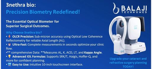
3NETHRA BIO - Optical Biometer
👇 Additional Feature 👇
COMPREHENSIVE MYOPIA MANAGEMENT REPORT AVAILABLE
Send your query for more information.