DIGITAL FUNDUS CAMERA
OVERVIEW
A Digital Fundus Camera is a specialized ophthalmic imaging device designed to photograph the interior surface of the eye, including the retina, optic disc, macula, and posterior pole. These high-resolution retinal images are essential for the early detection, diagnosis, and monitoring of various ocular and systemic diseases.
What It Does
A digital fundus camera allows for:
- High-resolution imaging of the retina and internal eye structure
- Non-invasive documentation of eye health over time
- Early detection of sight-threatening conditions, including:
- Diabetic Retinopathy
- Age-related Macular Degeneration (AMD)
- Glaucoma
- Retinal Detachment
- Hypertensive Retinopathy
How It Works
Light Source: Illuminates the retina via flash or continuous light.
Camera Lens System: Focuses on internal eye structures through the pupil:
Digital Sensor: Captures the image (typically using CCD or CMOS technology):
Software Processes, displays, stores, and optionally analyzes the captured images.
Types of Digital Fundus Cameras
Mydriatic: Requires pupil dilation for clearer, more detailed imaging.
Non-mydriatic: Works without dilation—faster, more comfortable, ideal for screenings.
Handheld/Portable Compact, battery-powered options for fieldwork or remote locations.
Ultra-Widefield Captures up to 200° of the retina in a single shot (vs. 30–45° standard).
Advanced Features & Integration
Modern digital fundus cameras may include:
- AI-Powered Analysis – Automated detection of diabetic retinopathy, AMD, glaucoma, etc
- Telemedicine-Ready – Share images remotely with specialists or consultants
- EMR Integration – Streamlined image upload into electronic medical records
- Image Enhancement Tools – Improve clarity for better diagnostic accuracy
- Cloud Storage – Secure access and archival of patient imaging data
Common Use Cases
Routine Eye Exams: Baseline retinal imaging for healthy patients.
Diabetic Screening Programs: Early detection and monitoring of diabetic retinopathy.
Glaucoma Monitoring: Track optic nerve head and cup-disc ratio changes.
Surgical Documentation: Pre- and post-operative records for retinal surgeries.
Academic & Research Institutions Teaching, research, and case study documentation.
Copyright © 2017 Balaji Technomed - All Rights Reserved.
**Product may vary from images on the website.
THIS WEBSITE USES COOKIES.
“We use cookies to improve your browsing experience and analyze website traffic. By accepting, your browsing data will be combined with other users’ data to help us optimize our website.”

Exclusive Introductory Deal
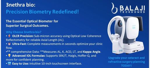
3NETHRA BIO - Optical Biometer
👇 Additional Feature 👇
COMPREHENSIVE MYOPIA MANAGEMENT REPORT AVAILABLE
Send your query for more information.